DIGESTION AND ABSORPTION
DIGESTION AND ABSORPTION
- Food is one of the basic requirements of all living organisms.
- The major components of our food are carbohydrates, proteins and fats.
- Vitamins and minerals are also required in small quantities.
- Food provides energy and organic materials for growth and repair of tissues.
- The water we take in, plays an important role in metabolic processes and also prevents dehydration of the body.
- Biomacromolecules in food cannot be utilised by our body in their original form. They have to be broken down and converted into simple substances in the digestive system.
- This process of conversion of complex food substances to simple absorbable forms is called digestion and is carried out by our digestive system by mechanical and biochemical methods.
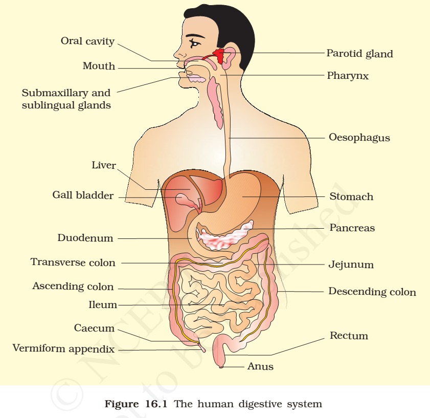
- General organisation of the human digestive system is shown in Figure 16.1.
16.1 DIGESTIVE SYSTEM
- The human digestive system consists of the alimentary canal and the associated glands.
16.1.1 Alimentary Canal
- The alimentary canal begins with an anterior opening – the mouth, and it opens out posteriorly through the anus.
- 30 feet is the length of alimentary canal in man.
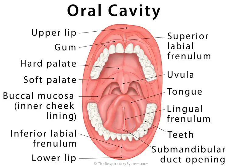
- The mouth leads to the buccal cavity or oral cavity.
- The oral cavity has a number of teeth and a muscular tongue. Each tooth is embedded in a socket of jaw bone.
- This type of attachment is called thecodont. Majority of mammals including human being forms two sets of teeth during their life, a set of temporary milk or deciduous teeth replaced by a set of permanent or adult teeth. This type of dentition is called diphyodont.
- An adult human has 32 permanent teeth which are of four different types (Heterodont dentition), namely, incisors (I), canine (C), premolars (PM) and molars (M).
- Incisors, Canines and lower Premolars have only one root.
- First premolars of upper jaw and molars of lower jaw has 2 roots.
- Second premolar may have 1 or 2 roots.
- all the molars of upper jaw has 3 roots.
- Arrangement of teeth in each half of the upper and lower jaw in the order I, C, PM, M is represented by a dental formula which in human is 2123/2123.
- Infant teeth 2102/2102
- The hard chewing surface of the teeth, made up of enamel, helps in the mastication of food.
The tongue:
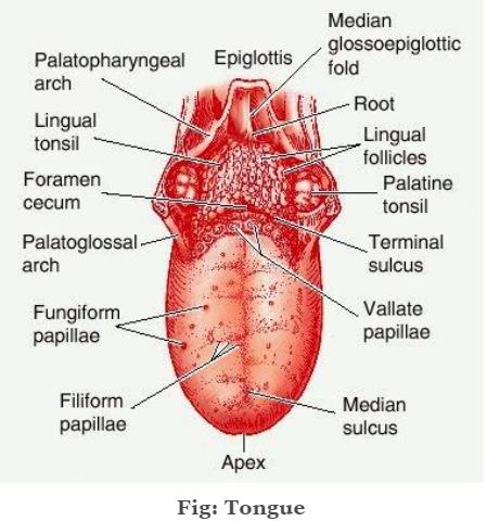
- The tongue is a freely movable muscular organ attached to the floor of the oral cavity by the frenulum.
- The upper surface of the tongue has small projections called papillae (filiform-sour, fungiform-sweet & salt, circumvallate-bitter), some of which bear taste buds.
- The oral cavity leads into a short pharynx which serves as a common passage for food and air.
- The oesophagus and the trachea (wind pipe) open into the pharynx.
- A cartilaginous flap called epiglottis prevents the entry of food into the glottis – opening of the wind pipe – during swallowing.
- The oesophagus is a thin, long tube which extends posteriorly passing through the neck, thorax and diaphragm and leads to a ‘J’ shaped bag like structure called stomach.
- A muscular sphincter (gastro-oesophageal) regulates the opening of oesophagus into the stomach.
- The stomach, located in the upper left portion of the abdominal cavity, has three major parts – a cardiac portion into which the oesophagus opens, a fundic region and a pyloric portion which opens into the first part of small intestine (Figure 16.3).

Small intestine:
- Small intestine is distinguishable into three regions, a ‘C’ shaped duodenum, a long coiled middle portion jejunum and a highly coiled ileum.
- The opening of the stomach into the duodenum is guarded by the pyloric sphincter.
- Ileum opens into the large intestine, through ilio-caecal valve.
The large intestine:

- It consists of caecum, colon and rectum.
- Caecum is a small blind sac which hosts some symbiotic micro-organisms.
- A narrow finger-like tubular projection, the vermiform appendix which is a vestigial organ arises from the caecum.
- The caecum opens into the colon.
- The colon is divided into three parts – an ascending, a transverse and a descending part.
- The descending part opens into the rectum which opens out through the anus.
- The wall of the alimentary canal from oesophagus to rectum possesses four layers (Figure 16.4) namely serosa, muscularis, sub-mucosa and mucosa.
- Serosa is the outermost layer and is made up of a thin mesothelium (epithelium of visceral organs) with some connective tissues.
- Muscularis is formed by smooth muscles usually arranged into an inner circular and an outer longitudinal layer.
- An oblique muscle layer maybe present in some regions. The submucosal layer is formed of loose connective tissues containing nerves, blood and lymph vessels.
- In the duodenum, glands are also present in sub-mucosa.
- The innermost layer lining the lumen of the alimentary canal is the mucosa.
- Mucosa layer forms irregular folds (rugae) in the stomach and small finger-like foldings called villi in the small intestine.
Villi:
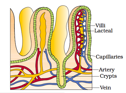

- The cells lining the villi produce numerous microscopic projections called microvilli giving a brush border appearance.
- These modifications increase the surface area enormously.
- Villi are supplied with a network of capillaries and a large lymph vessel called the lacteal.
- Mucosal epithelium has goblet cells which secrete mucus that help in lubrication.
- Mucosa also forms glands in the stomach (gastric glands) and crypts in between the bases of villi in the intestine (crypts of Lieberkuhn).
- All the four layers show modifications in different parts of the alimentary canal.


Comments
Post a Comment